Blood Vessels Labeled Brain - Chapter 19 blood vessels : He says the restricted vessels prevent the blood from draining fast enough and injure the brain by causing a build up of iron which leads to ms.
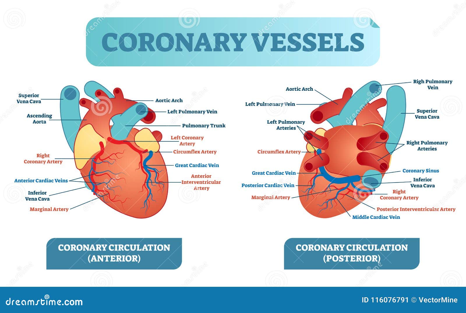
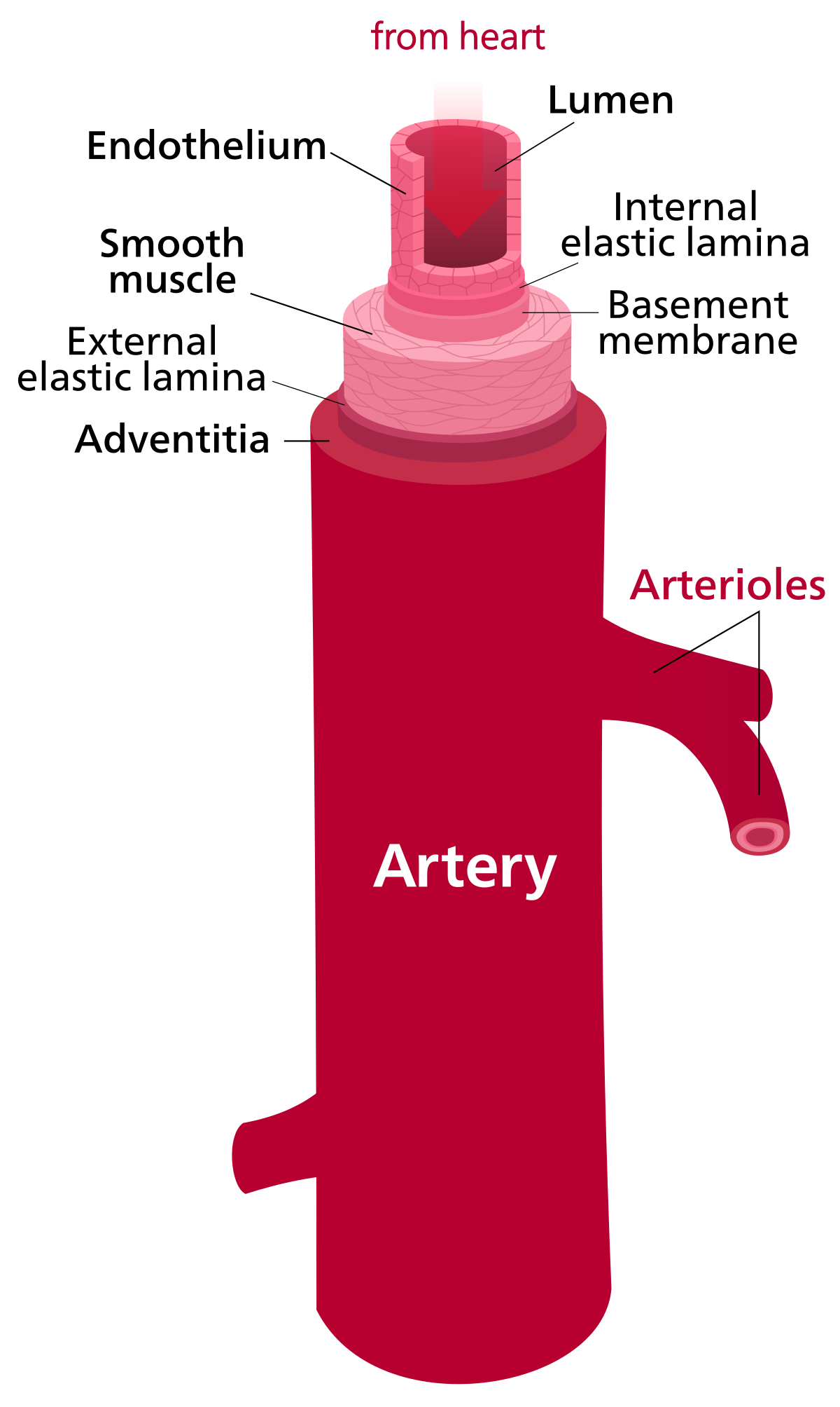
Blood Vessels Labeled Brain - Chapter 19 blood vessels : He says the restricted vessels prevent the blood from draining fast enough and injure the brain by causing a build up of iron which leads to ms.. Blood travels from the heart in arteries, which branch into smaller and smaller vessels, eventually becoming arterioles. The blood vessel wall is endowed with connective tissue, smooth muscle, and striated muscles. Only some of the vessels that exist in a real brain have been labeled. This is particularly important structure due to its clinical implications, which are discussed in more detail in the article. In the article on the ventricles within the cns, we will discuss their structure and.
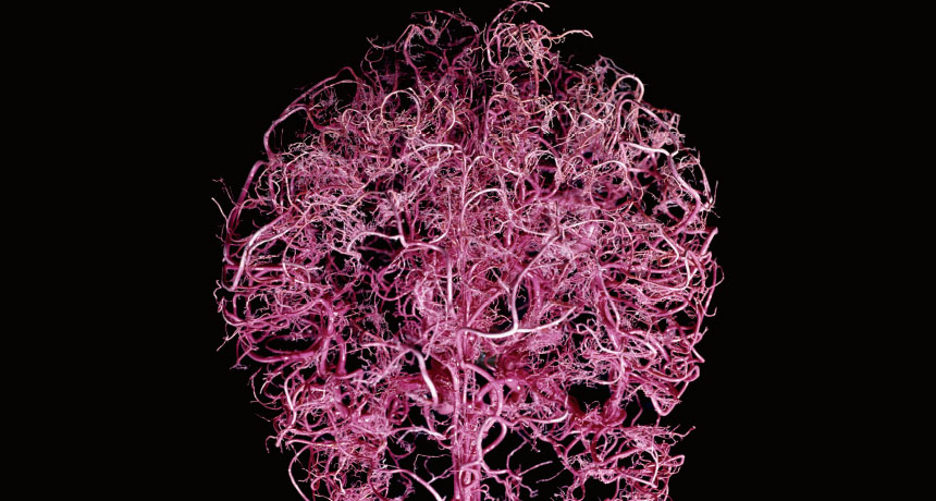
As well as providing new insights into the. The blood vessels in the brain are different, perhaps less willing to allow large molecules through the blood vessel walls. The blood vessels (and nerves) enter the brain through holes in the skull called foramina. Blood vessels in red in close communication with proliferating neuronal cells in the mouse cortex at embryonic day 10. Posterior communicating a internal carotid а.
heart: Heart Veins And Arteries Labeled from thumbs.dreamstime.com As well as providing new insights into the. Function and homeostasis of the brain relies on communication between its complex network of cells. Cerebral arterial circle anterior communicating posterior cerebral a middle cerebral al reset zoom. Blood vessels are vital for the body and play a key role in diabetes helping to transport glucose and insulin. The blood vessels (and nerves) enter the brain through holes in the skull called foramina. Supplies the anterior brain and the vertebral a. Supplies the posterior brain, blood supply to the entire brain is ensured by anastomoses between the vessels. Another whole article within the blood vessels and csf section is dedicated to the cavernous sinus.
There is a right sided aca and a left sided aca.
Posterior communicating a internal carotid а. Another whole article within the blood vessels and csf section is dedicated to the cavernous sinus. Examine a second specimen and notice any differences, such as asymmetries in the size of the vertebral or posterior communicating arteries. The blood vessels in the brain are different, perhaps less willing to allow large molecules through the blood vessel walls. He says the restricted vessels prevent the blood from draining fast enough and injure the brain by causing a build up of iron which leads to ms. Cerebral arterial circle anterior communicating posterior cerebral a middle cerebral al reset zoom. As well as providing new insights into the. Blood travels from the heart in arteries, which branch into smaller and smaller vessels, eventually becoming arterioles. The two cell types ensure the integrity of the neural vasculature by maintaining the blood. However, they have observed blood vessel damage caused. In the article on the ventricles within the cns, we will discuss their structure and. Blood vessels in red in close communication with proliferating neuronal cells in the mouse cortex at embryonic day 10. Blood in the brain is supplied by two pairs of large blood vessels (arteries):
Blood vessels innervate all tissues in vertebrates, enabling their survival by providing the necessary nutrients, oxygen, and hormonal signals. They also take waste and carbon dioxide away from the tissues. • identification of blood vessels as arteries, capillaries or veins from the structure of their walls. Microscopically, it is formed by the endothelium of the blood vessel. Brain vessel segmentation is a fundamental component of cerebral disease screening systems.
Blood exerts a powerful influence on the brain | Science News from www.sciencenews.org Supplies the anterior brain and the vertebral a. Label the veins of the anterior forearm and hand. Blood vessels are intricate networks of hollow tubes that transport blood throughout the entire body so that it can deliver valuable nutrients to and remove waste from cells. The blood vessel wall is endowed with connective tissue, smooth muscle, and striated muscles. Equal to the intestinal muscles that move the food morsel along brain level: • identification of blood vessels as arteries, capillaries or veins from the structure of their walls. Blood vessels are referred to collectively as the vascular system and, together with the heart, make up the circulatory system or cardiovascular system. Posterior communicating a internal carotid а.
He says the restricted vessels prevent the blood from draining fast enough and injure the brain by causing a build up of iron which leads to ms.
Fill in the blanks with the appropriate words to describe blood flow from the heart. Supplies the anterior brain and the vertebral a. The blood vessels in the brain are different, perhaps less willing to allow large molecules through the blood vessel walls. Label the veins of the anterior forearm and hand. Another whole article within the blood vessels and csf section is dedicated to the cavernous sinus. Label the blood vessels in the inferior view of the brain using the hints provided. The dense tight junctions between endothelial cells prevent paracellular transport through the. The precise relation between blood vessels and brain regions, reflecting the physiology and pathology of brain function directly and accurately, has figure 3. Blood vessels are referred to collectively as the vascular system and, together with the heart, make up the circulatory system or cardiovascular system. Instead, transport is controlled mostly by the dilation of vessels. Brain vessel segmentation is a fundamental component of cerebral disease screening systems. Blood is also supplied to the brain by the vertebral a. Blood vessel endothelium is continuous with the inner tissue lining of organs such as the brain, lungs, skin, and heart.
Label the blood vessels in the inferior view of the brain using the hints provided. The precise relation between blood vessels and brain regions, reflecting the physiology and pathology of brain function directly and accurately, has figure 3. Label the blood vessels of the male pelvis using the hints provided. Posterior communicating a internal carotid а. • identification of blood vessels as arteries, capillaries or veins from the structure of their walls.
Artery - Wikipedia from upload.wikimedia.org These vessels transport blood cells, nutrients, and oxygen to the tissues of the body. Instead, transport is controlled mostly by the dilation of vessels. Fill in the blanks with the appropriate words to describe blood flow from the heart. The 500 ms patients, both adults and children, also underwent mri scans of the brain to measure iron deposits in surrounding areas of the brain. Brain vessel segmentation is a fundamental component of cerebral disease screening systems. They also take waste and carbon dioxide away from the tissues. Cerebral arterial circle anterior communicating posterior cerebral a middle cerebral al reset zoom. The difference in the structural characteristics of arteries, capillaries and veins is attributable to their respective functions.
Identify all of the blood vessels that are illustrated in the figure as you can while holding or otherwise examining whole brain specimens.
The difference in the structural characteristics of arteries, capillaries and veins is attributable to their respective functions. The dense tight junctions between endothelial cells prevent paracellular transport through the. The structure, distribution and labeling of the whole brain vascular system of different arteries and veins in 3d. Blood vessels innervate all tissues in vertebrates, enabling their survival by providing the necessary nutrients, oxygen, and hormonal signals. This vessel supplies blood to the front part of your brain, knows as your frontal lobe. The precise relation between blood vessels and brain regions, reflecting the physiology and pathology of brain function directly and accurately, has figure 3. Label the blood vessels in the inferior view of the brain using the hints provided. This is particularly important structure due to its clinical implications, which are discussed in more detail in the article. Function and homeostasis of the brain relies on communication between its complex network of cells. The 500 ms patients, both adults and children, also underwent mri scans of the brain to measure iron deposits in surrounding areas of the brain. Examine a second specimen and notice any differences, such as asymmetries in the size of the vertebral or posterior communicating arteries. However, detecting vessels is still a challenging task those labeled as background or vessel voxels are excluded from consideration in later computation. Supplies the posterior brain, blood supply to the entire brain is ensured by anastomoses between the vessels.
The carotid arteries and the vertebral arteries anterior cerebral artery (aca): blood vessels labeled. The blood vessels (and nerves) enter the brain through holes in the skull called foramina.


0 Komentar